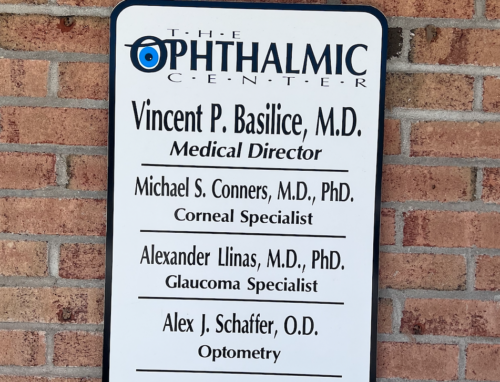
Meet the Doctors at TOC Eye
Whether you’re looking for cataract surgery, tips on how to manage your glaucoma, or you simply want new glasses, our team of award winning eye care specialists are ready to provide professional, personalized care.
At TOC Eye, our goal to provide exceptional eye care that’s tailored to meet the unique needs of each patient. We blend cutting-edge technology with personalized attention to deliver comprehensive eye exams, precise surgical procedures, and advanced treatments for a wide range of ocular conditions. Whether you require routine eye care, glaucoma management, cornea treatment, or are interested in learning more about LASIK surgery, our state-of-the-art facility is ready to address all your visual needs. Discover a clearer, brighter world with TOC Eye, where your vision is our top priority.

Cataracts are one of the most common vision issues in people over the age of 60. By undergoing cataract surgery, and with the help of several premium lens options, you can restore your vision and enhance your quality of life. The award-winning surgeons at TOC Eye will work with you to find the right lens to fit your needs and walk you through every step of the process so you can feel comfortable and confident about your procedure.
Learn More About Cataracts
Laser Vision Correction Surgery or LASIK is the world’s most popular vision correction procedure. LASIK is a safe, quick and effective way to improve your vision with little to no dependence on glasses and contact lenses. If you are frustrated with the constant maintenance, costs and inconvenience of glasses and contact lenses, LASIK may be for you. More than 40 million LASIK procedures have been performed to date making it the most common elective vision procedure in the United States.
Find out if you’re a LASIK candidate and take the next steps on your journey to better vision!
Take the LASIK Self TestWhether you’re looking for cataract surgery, tips on how to manage your glaucoma, or you simply want new glasses, our team of award winning eye care specialists are ready to provide professional, personalized care.
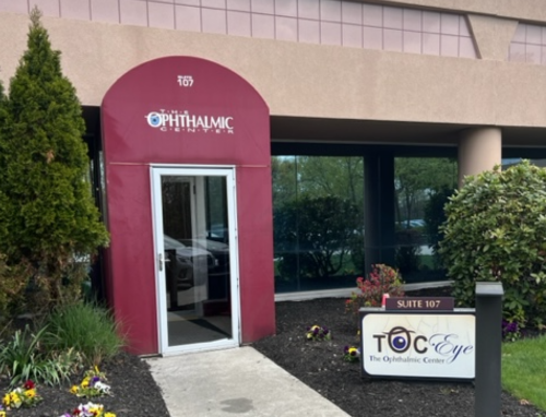
We strive to provide the highest quality eye care to the people in our communities. With offices across the north shore of Suffolk County, Long Island, the doctors at TOC Eye are here for you when you need them most.
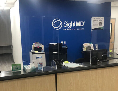
Looking for a specialty that’s not offered at East Setauket or Wading River? TOC Eye patients have access to all services at all SightMD locations.
Our top doctors are here for you to answer all your questions and help you find the right surgery options to fit your lifestyle. Fill out the form below to schedule your cataract consultation, our team is looking forward to hearing from you!